Epitope mapping before design: how do you choose which site to target?
Epitope mapping answers where an existing antibody binds. Designing one reverses the question: you choose the site first. How we read a target surface, which sites are worth engaging, which are not, and what that decision costs if you get it wrong.

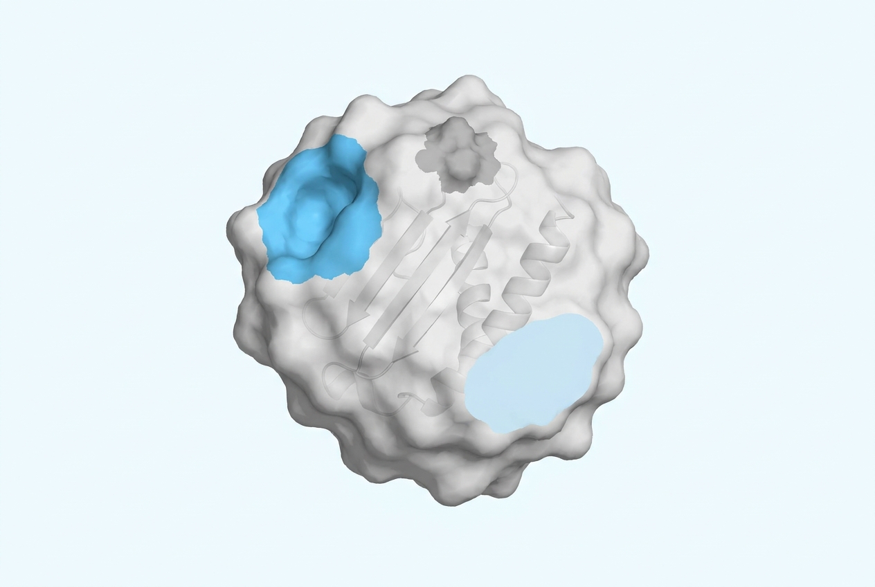
Epitope mapping normally answers a question about a molecule you already have: where on the antigen does this antibody bind? When the antibody is going to be designed rather than discovered, the question runs the other way. You choose the site first, and the design follows. That reversal is recent, it is the consequence of being able to specify an epitope computationally, and it moves the hardest decision in the project to the very beginning. A good binder against the wrong site is a failed programme, and it fails late.
Epitope mapping and epitope choice are two different jobs
Classical epitope mapping is a measurement. You have a binder, and you want to know what it touches. X-ray crystallography or cryo-electron microscopy of the complex gives you the contact residues directly. Hydrogen-deuterium exchange mass spectrometry gives you the protected regions. Alanine or shotgun mutagenesis gives you the residues whose substitution kills binding. Epitope binning, a related exercise, sorts a panel of antibodies into groups that compete with each other, telling you how many distinct sites your panel covers without telling you where they are.
These are experimental services, and Immunochem does not provide them. What we do is the other half: reading a target surface before any binder exists, and deciding which site is worth engaging.
The two jobs also need different inputs. Mapping requires a purified antigen and a binder to measure, whereas choosing requires a structure, a biological hypothesis about what the binder has to accomplish, and a judgement about which surfaces are tractable.
Before epitope mapping, settle what the binder has to do
Nothing about site selection makes sense until this is settled, and it is settled by biology rather than by structure.
- Block an interaction: the binder has to overlap the interface the antigen uses, so the site is defined by the partner protein rather than by whichever surface looks convenient. Where a co-structure of the complex exists, the decision is largely made for you.
- Inhibit an enzyme: the active site or an allosteric pocket, both usually recessed, which is where a convex single-domain paratope has the advantage over a flat one, as set out in our note on what a nanobody is.
- Detect or image without perturbing: here the requirement is almost reversed. You want a site that is accessible, abundant, conserved across the variants you need to detect, and functionally inert, so that binding reports presence without altering behaviour.
- Discriminate between two similar proteins: the site has to contain the residues that differ between them. This constraint is severe and frequently decides whether the project is viable at all, since those discriminating residues may sit on a surface that is otherwise poor for binding.
- Stay clear of someone else's claims: a constraint of its own, covered in our note on designing around an antibody patent. It bears on site choice directly whenever the claim follows the binding site instead of the sequence.
Six properties that separate a designable site from a poor bet
Given the functional requirement, several surfaces will usually satisfy it, and they are rarely equally designable. These are the properties that separate them:
- Shape: concave and pocketed sites give a paratope something to grip and suit an extended CDR3 loop particularly well. Flat, featureless surfaces are the hardest, because there is little to bind beyond chemistry.
- Chemistry: a mixed surface carrying hydrophobic patches and a few hydrogen-bonding anchors is what a design algorithm can exploit. Uniformly polar or highly charged surfaces are considerably harder, and heavily glycosylated regions are usually unavailable altogether, since the glycan occupies the space and varies with the expression system.
- Rigidity: a site that holds a single conformation can be designed against, whereas a disordered loop cannot, because the structure you designed against is only one of many that the region adopts. This is the most common reason a computationally attractive site may turn out badly in the laboratory.
- Conservation: where the binder has to work across species for a toxicology model, or across variants of a pathogen, the site must be conserved in all of them. Checking this costs an alignment and saves months.
- Uniqueness: a site with close homologues elsewhere in the proteome is a specificity problem waiting to happen. Off-target binding to an unrelated human protein is one of the expensive late failures described in our note on why antibody programs fail.
- Prior evidence: a site that a published antibody already engages should be read as encouraging, since somebody has demonstrated both that the surface supports binding and that engaging it has an effect. The constraint it introduces is legal rather than technical.
What the free feasibility study assesses, and what it returns
The input is a structure. An experimental structure where one exists, a predicted model otherwise, and a co-structure with a known partner or antibody where that is available, since it decides the question rather than informing it.
We then rank the candidate surfaces on the properties above, score how designable each one is, and return that assessment with our reading of which site best serves what you are trying to do. Where the functional requirement already fixes the site, the work is narrower: we assess whether that specific site is one we can credibly design against, and we say so when it is not.
What choosing the wrong site costs, stage by stage
Site selection is cheap to do and expensive to get wrong.
A disordered loop yields designs that bind the model rather than the protein. A glycan-covered surface yields nothing that binds at all. A conserved site chosen where a discriminating one was needed yields a binder that cannot tell your two proteins apart, which you discover during specificity testing once the molecule already exists. And a functionally inert site chosen where a blocker was needed yields a reagent instead of an inhibitor.
In each of those cases the campaign fails even though the design work was carried out competently, and the failure surfaces at the validation stage, after the design work, the synthesis and the assay development have all been paid for.
Set against that, site selection amounts to a day or two of structural analysis, and the asymmetry between the two is the whole argument for making the choice deliberately.
Receive a feasibility study on your antigen - for free
Frequently asked questions
What is epitope mapping?
Epitope mapping determines which residues on an antigen an antibody contacts. It is done experimentally, by structural methods such as X-ray crystallography and cryo-electron microscopy, by hydrogen-deuterium exchange mass spectrometry, or by mutagenesis scanning. It requires an antibody that already exists.
What is the difference between epitope mapping and epitope binning?
Mapping locates the site. Binning sorts a panel of antibodies into competition groups, telling you how many distinct sites the panel covers without identifying where those sites are. Binning is faster and is often run first on a large panel, with mapping reserved for the leads. Both belong to a discovery workflow, where the binders already exist and their site is worked out afterwards. When the binder is designed instead, the order reverses: you choose the site first and the sequence is built against it.
Can you choose an epitope before the antibody exists?
Yes, and this is what computational design changed. Designing against a user-specified epitope with confirmed binding pose has been demonstrated experimentally (Bennett et al., Nature 2026, 649:183-193, DOI, reference located through PubMed). With immunisation you take the epitopes the immune system selects. With design you name the site and the campaign is aimed at it, and whether the binder engages that site has to be shown experimentally.
What makes an epitope undesignable?
Most often conformational flexibility: the site does not hold a single structure, so the model you designed against is not what the binder meets. After that, dense glycosylation, which blocks access, and large flat polar surfaces, which offer little for a paratope to exploit.
Do you need an experimental structure, or is a predicted model enough?
A predicted model is usually workable, and we say so when it is not. Confidence in the model matters more than its origin: a well-predicted rigid domain is a better starting point than a poorly resolved experimental structure of a flexible region.
Do you perform epitope mapping as a service?
No, but EVOBodies™ reads structures, assesses which sites are designable, and designs against the site you select. If what you need is the site chosen rather than measured, that is exactly the work we do, and the free feasibility study is where it starts.
Not sure which site on your target to go after?
Receive a feasibility study on your antigen - for free